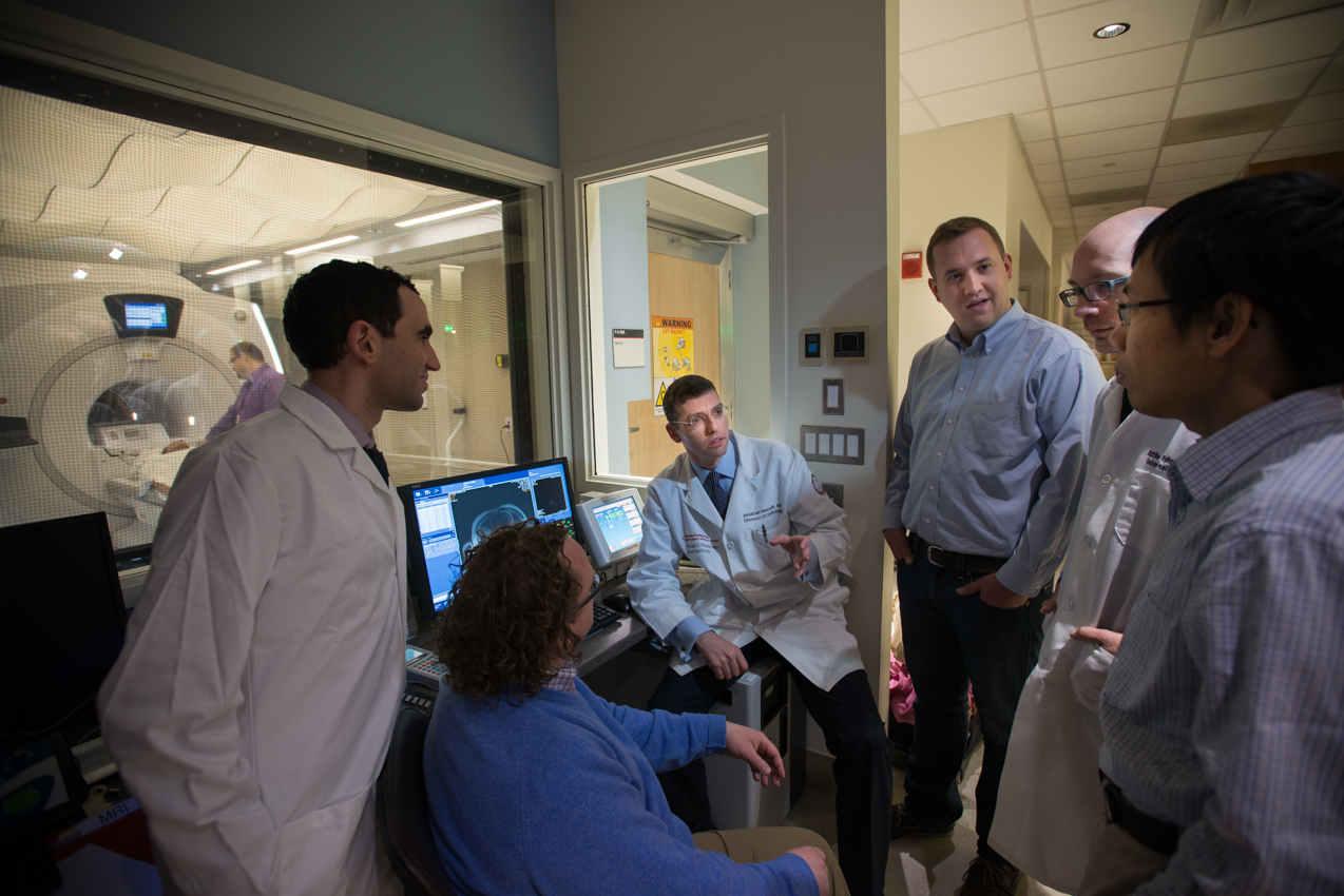
Mitral regurgitation (MR) is among the most common causes of valvular heart disease, affecting over 2 million people in the United States. Left untreated, MR, can cause serious consequences, including heart failure, arrhythmias, and even death. MR involves leakiness of one of the heart’s critical valves, the mitral valve, which causes the blood to flow backwards rather than forward. The good news is that there are effective treatments for MR, including valve replacement, angioplasty, bypass surgery, and medications. However, optimal treatment is dependent on determining actual causes for MR, which can result from problems with the mitral valve itself, as well as disorders within the supporting heart muscle. Using a new approach with a cardiac MRI, cardiologists are able to peer into the very tissues of the heart to assess the level of infarction (damage to the heart after a heart attack) at a vastly improved level compared to standard geometrical and function indices. Dr. Jonathan W. Weinsaft and colleagues are at the forefront of this new advance and their findings from a multidisciplinary study involving 150-plus patients were published in the JACC: Cardiovascular Imaging.
“Think high definition TV compared to standard TV,” says Dr. Weinsaft. “We now have a 3D view directly into the papillary muscles of the heart, allowing us to see tissue properties in the heart at a far higher resolution compared to standard approaches for MR tissue characterization. We can now review not only function and structure, but the tissue characteristic, or tissue properties, of the myocardium itself. We can now analyze the heart’s tissue, the papillary muscle itself, and better determine which patients will develop worsening MR versus patients who have MR that will get better on its own, and that does happen in some cases. MR is a dynamic process dependent on a complicated interaction of the heart muscle, the heart valve itself, and the structural tethering of the mitral valves. If it worsens, it can require a mitral valve replacement,”
The results that Dr. Weinsaft and colleagues have described in their paper represent a revolutionary change for the treatment of MR. “We are extending the paradigm,” explains Dr. Weinsaft, “with a new paradigm that not only looks at function and size of the heart, but looks specifically at tissue properties, namely the amount of infarction within the papillary muscles, as well as the left ventricular wall itself. We are applying standard MRI techniques to look for the amount of infarction in the heart but we are also extending this to apply new MRI techniques to determine which patients (following a heart attack) will develop substantial MR. As we move ahead with our studies, we will be better able to stratify which patients will respond to treatments like angioplasty or bypass surgery, based on the tissue properties within the myocardium.”
“The key thing,” says Dr. Weinsaft, “ is that there are different types of interventions and operations that will work for MR – a whole host of approaches – but now cardiologists can better assess how to link the disorder of the mechanism to a tailored treatment plan for the patient.”
Related Links:
Mitral Apparatus Assessment by Delayed Enhancement CMR (JACC: Cardiovascular Imaging)
Related Doctors:


