Echocardiology
Weill Cornell Medicine’s state-of-the-art Echocardiography laboratory, one of the busiest in the nation, performs over 30,000 studies a year. The Echocardiography lab at Weill Cornell is accredited under strict Intersocietal Accreditation Commission standards and offers a comprehensive spectrum of echocardiographic services and procedures, including Doppler, contrast, strain and 3-dimensionsal echocardiography; exercise and dobutamine stress echocardiography; transesophageal and intra-procedural echocardiography; and a wide array of vascular imaging.
Echocardiographic guidance is instrumental in guiding structural heart and electrophysiology procedures, such as transcatheter valve repairs and replacements, left atrial appendage occluders, and cutting-edge investigational devices.
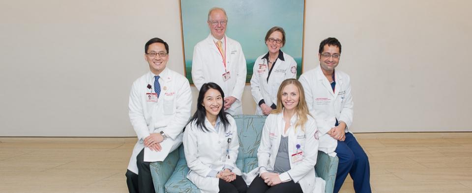
Our faculty members are leading experts in the field and have been involved in numerous clinical research studies. In addition, our faculty provide a structured imaging curriculum for our Cardiology fellows, which includes didactic lectures as well as intensive hands-on training in the performance and interpretation of echocardiograms.
Our faculty and laboratory have particular expertise in a number of areas including Marfan syndrome and aortopathy, valvular heart disease, adult congenital heart disease, and cardio-oncology.
All studies undergo meticulous assessment of the size and function of the heart, following standards established by our world-class research and contributions to national and international guidelines. Our detailed reports provide referring physicians and patients with a comprehensive evaluation of cardiac chamber dimensions; and systolic and diastolic function; valvular and adult congenital heart disease.
What is an Echocardiogram?
An echocardiogram (also referred to as an Echo or heart ultrasound) is a test that creates pictures of the heart using ultrasound imaging. Echocardiograms are safe, non-invasive diagnostic tests that work by using high pitched sound waves (not defected by the human ear), which are sent from a device called a transducer, and reflected off the different structures of the heart. These echoes are then turned into moving pictures of the heart that can be seen on a video or computer screen.
Echocardiograms are used to assess both the structure and the function of the heart. They can also be used to detect many types of heart disease and can also track the effectiveness of various medications and treatments.
Types of Echocardiograms
Transthoracic Echocardiography (TTE)
TTE is the most common type of echocardiogram. During this test, views of the heart are obtained by moving the Echo transducer to different locations on top of the chest wall or abdominal wall. Two-dimensional imaging of the heart is obtained, to assess the heart’s structure and function. In addition, using Doppler echocardiography, the direction and speed of blood flow can be checked as blood travels through the heart chambers, across the heart valves and through blood vessels.
Three-Dimensional Echocardiography
Three-dimensional echocardiography is a technique that uses advanced ultrasound imaging to create three-dimensional views of the heart, which provide more ways to see the heart chambers, heart function and heart valves.
Stress Echocardiography
A stress echocardiogram can be performed to look for narrowing in the arteries that supply blood flow to the heart muscle, which can suggest coronary artery disease (CAD). There are several other reasons to perform a stress echocardiogram, including heart conditions such as hypertrophic cardiomyopathy.
During a stress echocardiogram, ultrasound imaging is performed both before and after your heart is stressed. Exercise is a common method of stress for this procedure. For people who cannot exercise, medication-based stress testing can be performed. For example, dobutamine is a medication that is administered through a peripheral IV; it increases the pumping of the heart like during exercise.
Strain Echocardiography
Strain imaging can be performed during a standard echocardiogram. This technique is used to assess the heart muscle and its function.
Transesophageal Echocardiography (TEE)
TEE allows for more detailed views of the heart structures and their function. During a TEE, a thin flexible echo probe is passed down the esophagus, to provide closer pictures of the heart, and to avoid structures that can interfere with imaging such as the lungs and ribs. Before the TEE, a numbing spray is applied to the throat, and during the procedure an anesthesiologist provides sedation, to ensure comfort during the test.
Schedule an Appointment
To schedule an appointment for an echocardiogram please call (646) 962-4733. Your referring physician may also complete the Cardiac Echocardiography Lab Order Form.
Our Physicians
| Faculty | Title | Phone | ||
|---|---|---|---|---|
 |
Richard B. Devereux, M.D. |
Professor of Medicine, Director Adult Echocardiography Laboratory | 646-962-4733 | Full Profile |
 |
Eirini Apostolidou, M.D. |
Assistant Professor of Medicine | 646-962-4733 | Full Profile |
 |
Rebecca Ascunce, M.D. |
Assistant Professor of Medicine, Patient Safety & Quality Leader | 646-962-5558 | Full Profile |
 |
Sean Chen, M.D. |
Assistant Professor of Medicine | 646-962-5558 | Full Profile |
 |
Jennifer Chen, M.D. |
Assistant Professor of Medicine, Director Clinical Echo Operations, Director Nuclear Lab | 646-962-4733 | Full Profile |
 |
Stephanie Feldman, M.D. |
Assistant Professor of Medicine, Director Cardio-Oncology | 646-962-5558 | Full Profile |
 |
Sandeep Gangireddy, M.D. |
Assistant Professor of Clinical Medicine | 646-962-5558 | Full Profile |
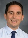 |
Edwin Homan, M.D. Ph.D. |
Assistant Professor of Medicine | 646-962-5558 | Full Profile |
 |
Jennifer K. Jantz, M.D. |
Assistant Professor of Medicine, Director Structural Heart Imaging | 646-962-4733 | Full Profile |
 |
Robert J. Kim, M.D. |
Associate Professor of Clinical Medicine, Director Consultative Cardiology | 646-962-5558 | Full Profile |
 |
Jiwon Kim, M.D. |
Associate Professor of Medicine, Director, Cardiovascular Imaging Program | 646-962-4733 | Full Profile |
 |
Samuel M. Kim, M.D. |
Assistant Professor of Medicine, Director Preventive Cardiology | 646-962-5558 | Full Profile |
 |
Vinay Kini, M.D. |
Associate Professor of Medicine, Associate Program Director, Cardiovascular Disease Fellowship Program | 646-962-4733 | Full Profile |
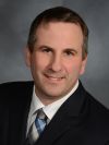 |
Saaron Laighold, M.D. |
Assistant Professor of Medicine, Director, Echocardiography Innovation | 646-962-5558 | Full Profile |
 |
Hannah Lehrenbaum, M.D. |
Assistant Professor of Medicine | 646-962-5558 | Full Profile |
 |
Julie L. (Friedman) Marcus, M.D. |
Assistant Professor of Clinical Medicine | 646-962-5558 | Full Profile |
 |
Alicia Mecklai, M.D. |
Assistant Professor of Medicine, Director 4 North Telemetry Service, Co-Director Women's Heart Program | 646-962-5558 | Full Profile |
 |
Nupoor Narula, M.D. M.S.c. |
Assistant Professor of Medicine, Director Cardiology Vascular Laboratory, Director Women's Heart Program | 646-962-4733 | Full Profile |
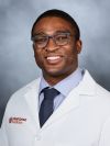 |
Chukwuma Onyebeke, M.D. |
Assistant Professor of Medicine | 646-962-5558 | Full Profile |
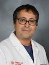 |
Harsimran Sachdeva Singh, M.D. |
Assistant Professor of Medicine, Director Adult Congenital Heart Disease, Director Cardiovascular Disease Fellowship | 646-962-2243 | Full Profile |
 |
Diala Steitieh, M.D. |
Assistant Professor of Medicine, Director Hypertrophic Cardiomyopathy | 646-962-5558 | Full Profile |
 |
Su Yuan, M.D. |
Assistant Professor of Clinical Medicine | 646-962-2243 | Full Profile |
 |
Robert Zhang, M.D. |
Assistant Professor of Medicine | 646-962-5558 | Full Profile |


