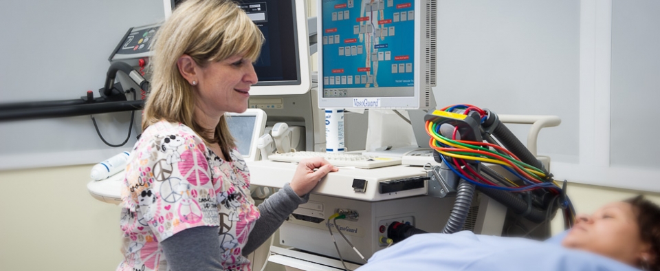
Vascular Imaging and Testing
Vascular imaging is a non-invasive test that evaluates blood vessels to determine if blood is flowing throughout your body in a healthy way. To accurately diagnose abnormalities in the vascular system, our specialists use the most advanced, contemporary noninvasive tests available. Vascular imaging is most often used to diagnosis vascular disease.
Our Services
Our state-of-the-art vascular laboratory is accredited by the Intersocietal Accreditation Commission for Vascular Testing, which means our lab meets the highest standards of quality patient care and monitoring. Annually, our lab performs over 2,000 vascular studies and offers the full spectrum of non-invasive diagnostic modalities for the detection of vascular disease. Some of our lab’s services include:
Measurement of the Ankle-Brachial Index with Pulse Volume Recordings
This painless, non-invasive method is often used as the initial screening for peripheral artery disease (PAD). Using standard blood pressure cuffs and a special stethoscope, blood pressure is measured in the arms and legs to determine how well blood flows through the vessels.
A treadmill exercise test may also be performed. During this test, the patient walks on a treadmill until a symptom appears. Once this happens, the blood pressures are measured again. This test is useful to determine the severity of the blockages in the leg arteries and may also be used to differentiate symptoms of vascular disease from other causes of leg pain.
Vascular Duplex Ultrasound Testing
This noninvasive procedure uses ultrasound to produce images of a blood vessel and to provide information on the speed with which blood travels through the vessel. Our lab offers the most advanced ultrasound testing for vascular disease, including:
- Arterial duplex ultrasonography for detecting abdominal aortic aneurysms as well as narrowing of the carotid arteries, renal arteries, and arteries of the arms and legs.
- Graft surveillance for those who have been treated for peripheral artery disease with a graft or stent. Ultrasound evaluation of the treated area is part of the follow-up procedure. This is done to ensure the graft remains patent and to try to identify any potential problems before they occur.
- Venous duplex imaging to detect blockages/blood clots in the veins of the legs.
- Venous ultrasonography of the upper and lower extremities.
Your providers may also refer you for additional testing, including computed tomography angiography or magnetic resonance angiography.
Angiograms for Further Evaluation
If obstructive vascular disease has been identified with these non-invasive tests, your physician may recommend an angiogram. An angiogram is an invasive test that is performed in the cardiac catheterization laboratory. A thin tube, called a catheter, is inserted into the groin and is led to the affected site. A dye is then introduced through the tube to allow the blockage to be visualized and evaluated prior to treatment.
Request an Appointment
Often, imaging studies and vascular and cardiac testing will require a referral from your physician and, in some cases, pre-authorization from your insurance company. Our patient coordinators are skilled in this process and will assist you every step of the way. To request an appointment for vascular imaging, please call (646) 962-4733.
Our Physicians
| Faculty | Title | Phone | ||
|---|---|---|---|---|
 |
Richard B. Devereux, M.D. |
Professor of Medicine, Director Adult Echocardiography Laboratory | 646-962-4733 | Full Profile |
 |
Samuel M. Kim, M.D. |
Assistant Professor of Medicine, Director Preventive Cardiology | 646-962-5558 | Full Profile |
 |
Nupoor Narula, M.D. M.S.c. |
Assistant Professor of Medicine, Director Cardiology Vascular Laboratory, Director Women's Heart Program | 646-962-4733 | Full Profile |
 |
Mary J. Roman, M.D. |
Professor of Medicine | 646-962-4733 | Full Profile |


