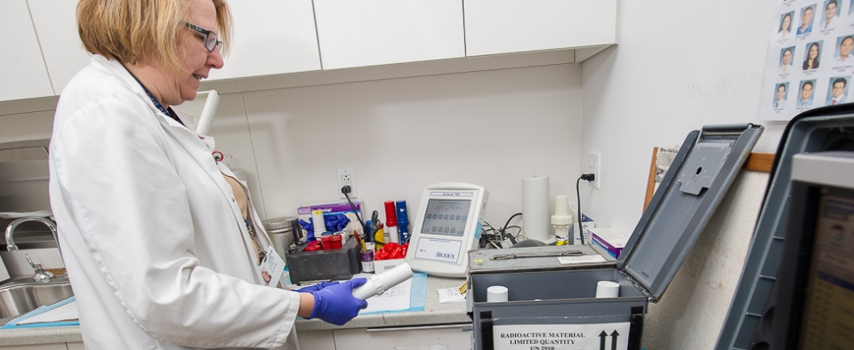
Nuclear Cardiology
Weill Cornell Medicine’s nuclear cardiology lab performs over 3,000 nuclear cardiac imaging studies annually and offers a variety of non-invasive cardiovascular imaging studies. Some of these studies include myocardial perfusion imaging (nuclear stress tests) for the detection and assessment of coronary artery disease, viability studies to assess for the extent of myocardial infarction, amyloid (PYP) scans, and radionuclide cineangiograms (RNCA/MUGA studies) to evaluate global heart function. In addition, stress modalities routinely performed include exercise treadmill testing and vasodilator chemical or pharmacologic stress (utilizing mostly regadenoson, dipyridamole, or dobutamine in patients who are unable to exercise).
Our nuclear cardiology lab uses cutting-edge solid-state cadmium zinc telluride (CZT) cameras to acquire a complete study in half the time (3-5 minutes) compared to conventional imaging protocols (15-20 minutes), and allow for a low amount of tracer. Our state-of-the-art facility is also accredited by the Intersocietal Accreditation Commission (IAC/Nuclear). Scans are performed with the highest level of sophisticated protocols that afford the most accurate assessment of blood flow to the heart and overall function while optimizing patient satisfaction, comfort, and care.
What is Nuclear Cardiology Imaging Testing?
Nuclear imaging evaluates how organs function, unlike other imaging methods that assess how organs appear. A small and safe amount of a radioactive tracer solution(s) are introduced into the body via an intravenous injection. A special camera detects the tracer solution in different parts of the body and a computer generates a series of images of the heart.
Nuclear cardiac imaging can help determine if there is adequate blood flow to the heart muscle during stress versus rest. This high-level imaging also accurately evaluates heart function and the presence of prior heart attacks.
Nuclear cardiac images help to identify coronary heart disease, the severity of prior heart attacks, and the risk of future heart attacks. This clinically important evaluation of cardiac function and amount of heart muscle at risk enable cardiologists to practice precision medicine, by refining prescription of medications and better selecting which patients need further testing such as coronary angiogram, the need for angioplasty or bypass surgery, or devices to optimize treatment outcomes.
Types of Nuclear Cardiology Imaging Testing?
Cardiac SPECT (Single Photon Emission Computed Tomography): Cardiac SPECT imaging, also called myocardial perfusion imaging, are non-invasive tests used to assess the heart’s structure, function and blood flow. A SPECT scan is obtained after an individual has completed either exercise on a treadmill or a chemical/pharmacologic stress test.
SPECT scans use small amounts of radioactive substances that are injected into a vein and a special camera produces images of the heart. These images allow Cardiologists to assess blood flow to the heart muscle and detect areas of abnormal heart muscle. Information obtained from SPECT scans can be used to:
- Identify blockages in the coronary arteries (blood vessels of the heart)
- Determine whether an individual has had a prior heart attack
- Predict which patients are at high risk for a future heart attack
- Assess the heart’s condition after cardiac bypass surgery or angioplasty (i.e. coronary artery stent placement)
Viability Imaging: Viability imaging is performed solely at rest (no stress component) and with a thallium tracer, and is used to evaluate blood flow to the heart. After a heart attack, a portion of the heart muscle may be irreversibly damaged. However, other regions of the heart may be injured, but not irreversibly damaged. These areas are referred to as hibernating. These hibernating regions of the heart muscle are not discernable with traditional imaging studies such as an echocardiogram. The injured heart regions may be viable, but have not fully healed. As a consequence, heart function may be temporarily reduced.
A viability study would identify hibernating regions and better inform your Cardiologist whether restoring blood flow to that region of the heart may improve heart function.
Amyloid Pyrophosphate (PYP) Imaging: Cardiac amyloidosis remains an underdiagnosed cause of heart failure or weakening of the heart muscle. There are two forms of amyloid deposits that may affect the heart: misfolded light chain or TTR (transthyretin) proteins. Differentiating these two forms of cardiac amyloid is vital in determining prognosis and treatment.
Imaging with our SPECT cameras utilizing a tracer, Tc-99m PYP, is highly accurate to noninvasively identify patients with TTR-type cardiac amyloid with a sensitivity and specificity of greater than 97%. This test is particularly instrumental in patients whom invasive myocardial biopsy is associated with unacceptable risk.
MUGA (Multiple Gated Acquisition) Scan: MUGA scan, also called radionuclide cineangiograms (RNCA), is a test that is used to evaluate heart function by measuring how much blood is pumped out of the ventricles of the heart with each heartbeat (ejection fraction). A small amount of a safe radioactive tracer solution is introduced into a vein. This substance attaches to red blood cells, which are visualized by a special camera and computer as they travel through the heart. The ejection fraction is then calculated based on the computer-generated images.
Schedule an Appointment
To schedule an appointment with nuclear cardiology please call (646) 962-5558 and have your referring physician complete the Nuclear Cardiology Lab Order Form.
Our Physicians
| Faculty | Title | Phone | ||
|---|---|---|---|---|
 |
Jonathan W. Weinsaft, M.D. |
Chief, Greenberg Division of Cardiology, A.M. Gotto Professor of Medicine, Atherosclerosis and Lipid Research | 646-962-5558 | Full Profile |
 |
Jennifer Chen, M.D. |
Assistant Professor of Medicine, Director Clinical Echo Operations, Director Nuclear Lab | 646-962-4733 | Full Profile |
 |
Sean Chen, M.D. |
Assistant Professor of Medicine | 646-962-5558 | Full Profile |


