

You may qualify for this study if you:
Are 18 years of age or older
Have advanced tricuspid regurgitation (TR) documented on a prior echocardiogram
See the full eligibility details below.
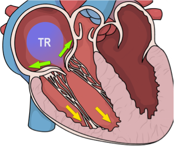
Tricuspid regurgitation (TR) is one of the most common forms of valvular heart disease in the United States and worldwide. Nearly 2 million Americans live with advanced TR that isn't caused by a structural problem with the valve itself (known as “functional TR”). As the population ages and conditions like coronary artery disease and heart failure become more common, the impact of functional TR is expected to grow substantially.
TR severity has a major effect on prognosis: patients with advanced TR face a 15% annual mortality rate, and five-year mortality reaches 65% among those with severe TR and reduced heart function. Research from our team and others has also shown that TR doubles the risk of symptomatic heart failure, with symptoms and exercise capacity closely tracking TR severity.
Despite how common and serious TR is, we still don't fully understand what drives poor outcomes in individual patients. Cardiac MRI gives us a unique window into this question. It can assess TR severity, right ventricle function and shape, and, importantly, the health of the heart muscle tissue near the tricuspid valve. Echocardiogram data adds a second perspective on TR and right ventricle changes and may help us develop simpler, more widely available tools informed by MRI to predict how patients will do over time.
Our goal is to identify new, tissue-based markers that can predict how functional TR will progress and how patients will respond to treatment. Findings from this study could shape how doctors monitor TR, guide decisions about intervention, inform the design of new devices, and improve future clinical trials, ultimately improving care for millions of people at risk.
If you decide to join, you will complete two study visits, one year apart. At each visit, you'll have:
Both the cardiac MRI and echocardiogram are paid for by the study. There is no cost to you.
You will also receive a $100 debit card at each visit. Your imaging reports will be made available to your care team through Epic.
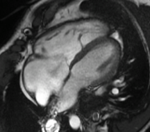
Cardiac MRI uses magnetic fields to create detailed pictures of your heart's shape and function. It also gives us information about the heart muscle itself, including blood flow and any signs of injury. MRI uses a contrast agent called gadolinium, which helps your doctors see the structures of your heart more clearly.
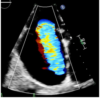
An echocardiogram (“echo”) uses sound waves (ultrasound) to create moving images of your heart as it beats. It's a routine, non-invasive test used to see how your heart is shaped and how well it's functioning.
You will not be eligible for this study if any of the following apply to you:
Participating in this study helps advance research that may improve how doctors understand and treat tricuspid regurgitation for you and for future patients. If you think you may be eligible and would like to learn more, our research team would be glad to answer your questions and explain what participation would involve.
Contact our research team:
Email: TRACT-TR@med.cornell.edu
Phone: 212-746-2627